


Welcome to the Micro Imaging Lab!
This lab is devoted to imaging natural materials at several scales, using 7 pieces of equipment:
(1) Photography station macro and micro, with a camera stand: This consists of a digital camera on an adjustable stand, with good lighting. This is especially useful for imaging large (5-50 cm) samples. Connection to a computer permits the user to photograph samples remotely, edit images, and save them digitally.
(2) Binocular and petro microscopes with digital cameras connected to a computer permit digital capture of close-up details.
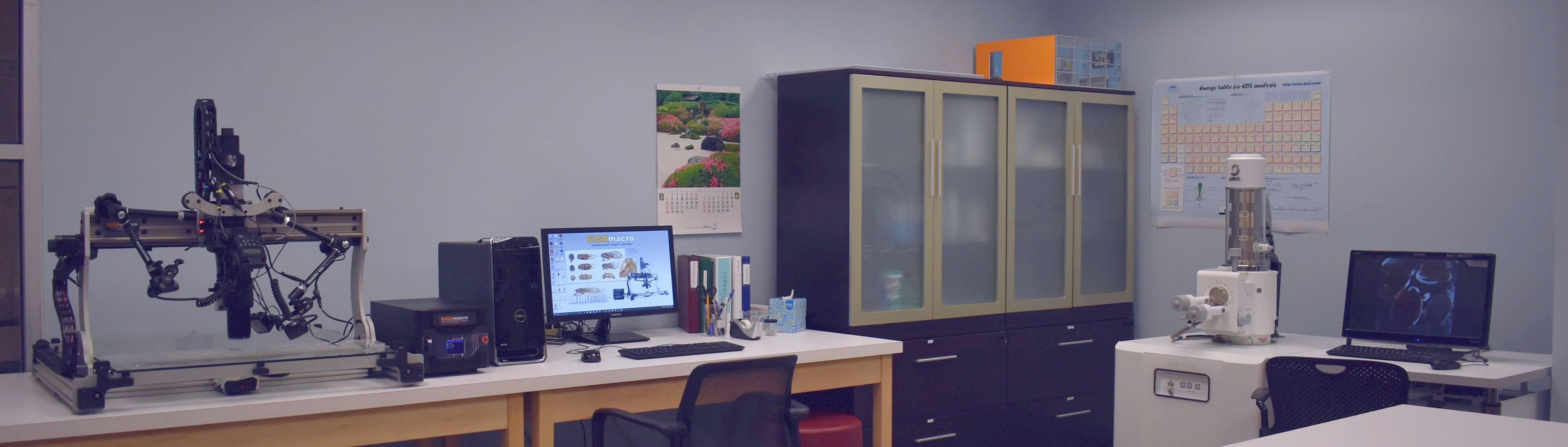
(3) GIGAmacro: This consists of a digital camera with zoom lens and computer software that combines focal stacking, image stitching, and 3D depthmaps for very high-resolution imaging of thin sections and other materials within a specific size range.
(4) JEOL Scanning Electron Microscope (SEM): Our SEM is flexible and powerful, using secondary electron imaging to show reveal very small surface topography, backscatter electron imaging to reveal phases with different mean atomic weight, and energy-dispersive X-Ray analysis of natural materials. The SEM is especially useful for studying regions that range in size from a millimeter to less than a micron.
(5) Thermo Niton XL3 Handheld XRF: This device analyzes the elemental composition of a hand sample in seconds using x-ray fluorescence and can be utilized for a wide range of applications, from art conservation to geochemistry and advanced materials science.
(6) ThermoFisher XRD Equinox 100
(7) ThermoFisher Raman Microscope
The lab is located at ROC 1.209b. Please contact Ignacio Pujana at pujana@utdallas.edu or Bob Stern at rjstern@utdallas.edu for more information. Use of the lab equipment is by appointment only.
NOTE: Our website is still under construction. Please check back often for new information and images.